In order to correctly follow any therapy, it is essentially important to know how to treat the structure.
The understanding of this structure is the basis of its function and significance in the entity of the body. This understanding would lead to the possibility of one correctly applying the relevant techniques to which it is applied.
In order to actualize a cranial technique, the osteopath is required to visualize the structure of the skull – according to Sutherland.
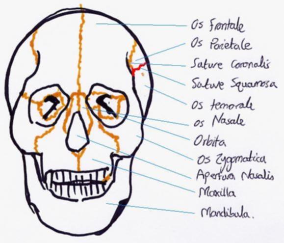
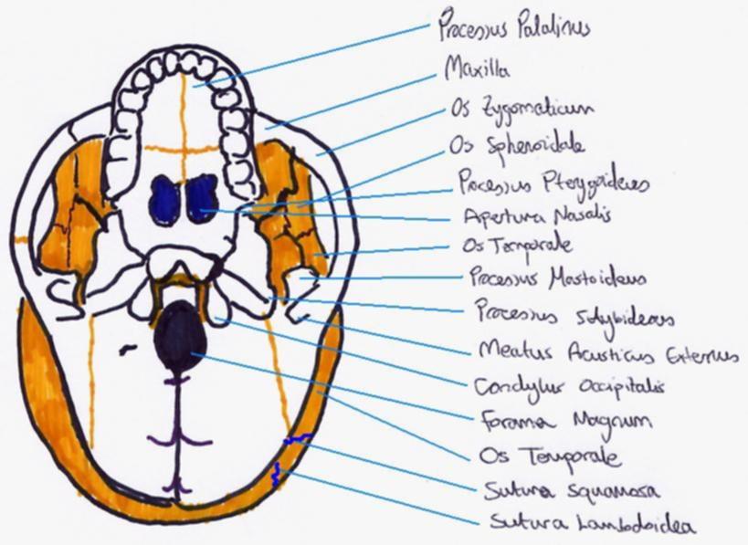
Descriptive Anatomy of the Cranial Bones
The skull is made up of 22 bones altogether, not including the bones used for
hearing.
The Cranial Bowl (Holding the brain):
1. Os Frontale - Non-pair. Forehead
Prenatal, exists as two bones. In 10% of cases, the sutures don’t join
between the two forehead bones. However, with the other 90% there is a high
malleability (flexibility) between the two halves – which can move as
two separate bones in the cranio-sacral rhythm.
2. Os Temporale - Pair. Temple bones
3. Os Parietal Crown bones
4. Zygomaticum - Pair. Cheek bones
5. Sphenoidal - Pair.
6. Occiput - Non-pair.
7. Ethmoid - Non-pair. Sieve bone
8. Vomer - Non-pair.
9. Os Nasale - Pair. Nasal bone
10. Os Lacrimale - Pair.
11. Concha Nasalis Inferior - Pair.
12. Mandibula - Non-pair. Lower Jaw
13. Maxilla - Pair. Upper Jaw
14. Os Palatinum - Pair. Palate Bone
Further bones : Os Hyoideum, Hearing bones.
Drawings